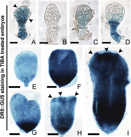
Fig. 4.
Auxin-dependent DR5::GUS expression in transformed embryos treated with TIBA in tobacco. (A–D) Abnormal early globular embryos from 3 DAP ovules after culture. (A, B) Abnormal globular embryos with asymmetrical division of EP and no apical–basal polarity, respectively. (C) An early globular embryo with faint GUS staining. (D) An abnormal embryo with deep GUS staining in the suspensor. (E) An abnormal undifferentiated embryo from the culture of 5 DAP ovules. (F–I) Abnormal differentiated embryos from the culture of 5 DAP ovules. (F) A cup-shaped embryo. (G–I) Abnormal differentiated embryos with two asymmetrical cotyledon primordia, three cotyledon primordia, and multiple cotyledons, respectively. Arrowheads indicate the division plane of EP cells in (A), cotyledon primordia in (H, I). (A–D) Bar=20 μm; (E) bar=40 μm; (F–J) bar=60 μm. (This figure is available in colour at JXB online.)

