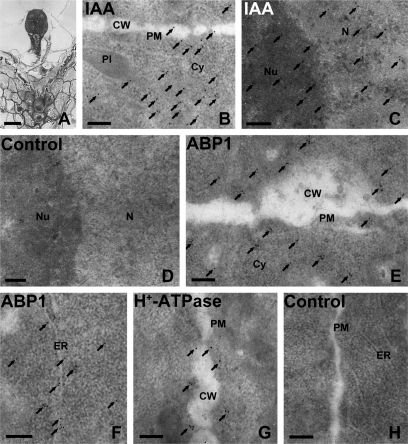
Fig. 9.
Immunogold localization of IAA, ABP1, and PM H+-ATPase in the early globular embryo (5 DAP) of tobacco. (A) Semi-thin section of an early globular embryo. (B–H) Ultrathin sections of the early globular embryo, showing IAA, ABP1, and PM H+-ATPase immunogold granules, respectively. (B) Numerous IAA gold particles appeared in the cytoplasm near the cell wall, and a few were distributed in the plasma membrane (PM) and cell wall of the embryo. (C) Many IAA gold particles accumulated in the nucleus. (D) A control section without the primary IAA antibody, and the secondary antibody was a goat anti-rat IgG antibody. The image showed no gold particles. (E, F) ABP1 gold particles mainly distributed in the PM and cytoplasm near the cell wall (E) and in the ER (F). (G) Nearly all PM H+-ATPase gold particles appeared in the PM. Arrows indicate the gold particles. (H) A control section without the primary antibodies of ABP1 or PM H+-ATPase, while the secondary antibody was goat anti-rabbit IgG antibody. The image showed no gold particles. (A) Bar=25 μm; (B) bar=5 μm; (C–E) bar=0.2 μm; Cy, cytoplasm; CW, cell wall; ER, endoplasmic reticulum; N, nucleus; Nu, nucleolus; Pl, plastid; PM, plasma membrane.

