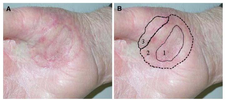
FIG. 2.

Patient 1. After 3 excisions and skin grafts (numbered and shown with solid, dashed, and dotted lines, respectively), there was still a positive margin at the radial aspect of excision 3. There is no clinically evident pigmented lesion. There is some fibrosis and scar from previous radiation and surgery.
