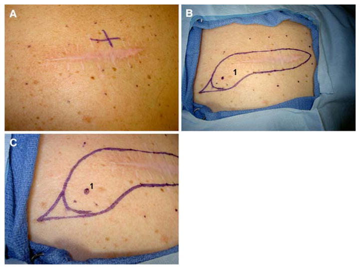
FIG. 4.

Patient 2. (A) Scar from previous melanoma excision with melanoma-in-situ (MIS) at multiple margins, multiple nevi, and extensive sun damage. (B) Mapping of biopsy sites, 1 and 2 cm away from the original scar and 1 cm apart from one another. Only biopsy 1 revealed any pathologic abnormality (MIS). (C) Magnified view of planned excision, including 1-cm margin around the scar and 1.5-cm margins around the positive biopsy site.
