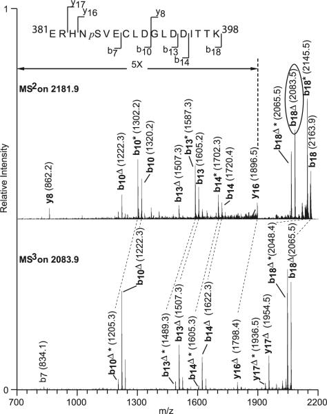
Fig. 4.
Tandem MS identifies S385 as a PKA phosphorylated residue. MS2 spectrum of 2,180.9-Da tryptic peptide fragments from the 625 U/μl PKA-treated GST-Kir3.1C. Fragmentation was carried out with 35% relative collision energy (Thermo's nomenclature). Subsequently, the phosphoric acid-depleted precursor ion b18Δ at m/z 2,083.9 (in oval) was selected for another fragmentation stage (MS3) using the same amount of collision energy. Ions from the b and y series are depicted indicating that the phosphorylated site is at serine 385. Dashed line indicates peak mass difference corresponding to loss of H3PO4; delta, loss of H3PO4 (98 Da); asterisk, neutral loss

