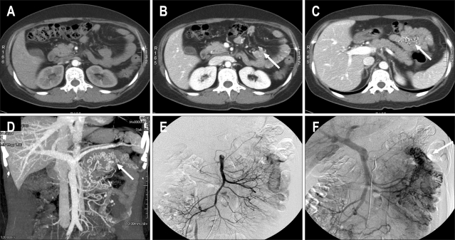
Fig. 1.
Axial CT image of the arterial phase showing no abnormal vessels. (B) Axial CT image of the portal phase showing enhancing abnormal veins at the antimesenteric border of the small bowel (arrow). (C) Axial CT image of the portal phase showing enhanced abnormal veins of the mesentery at the small bowel loop (arrow). (D) Maximum-intensity projection reconstruction image of CT angiography showing tortuous abnormal veins in the left upper abdomen (arrow). (E) Superior mesenteric artery (SMA) arteriography of the arterial phase showing no abnormal vessels. (F) SMA arteriography of the portal phase showing enhancing abnormal veins at the mesentery of the small bowel loop (arrow).

