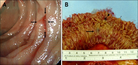
Fig. 2.
(A) Intraoperative endoscopy showing a 1.0-cm-diameter superficial ulcer covered with a blood clot that was 70 cm from the ligament of Treitz. (B) Gross examination revealed a 0.9-cm ulceration in the mucosa as a bleeding focus (arrowhead) and one blue-black lesion in the submucosal layer, with venous distension (arrow).

