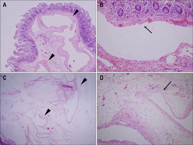
Fig. 3.
Microscopic images of the blue-black-colored polypoid submucosal lesion (A and B) and subserosal lesion (C and D). A and C: Ectatic thin-walled vessels with a few smooth muscle cells (arrowheads). B and D: Some foci of vascular walls were lined by only a single layer of endothelial cells (arrows) (H&E staining; A, ×40: B, ×200: C, ×12.5: D, ×200).

