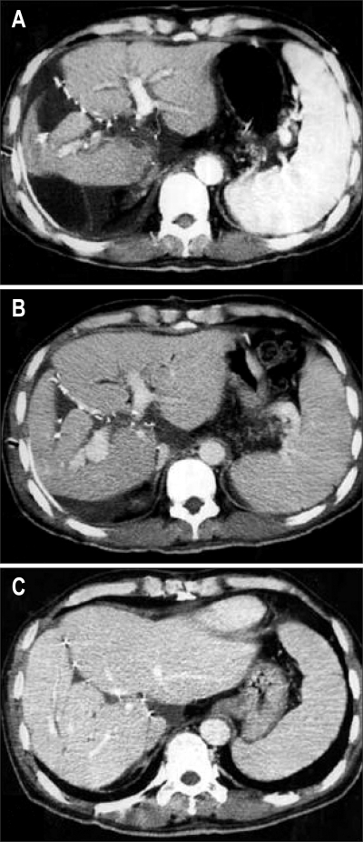
Fig. 14.
Postoperative follow-up CT scan of the recipient, demonstrating the balanced regeneration of both liver grafts. (A) CT scan taken 5 days after transplantation showing that the second left lobe graft in the right upper abdomen was still small and supported by a tissue expander bag. (B) CT scan made 2 weeks after transplantation, showing the rapid regeneration of both grafts. (C) CT scan made 2 months after transplantation, showing that two regenerated left lobe grafts were in the shape of a normal liver.

