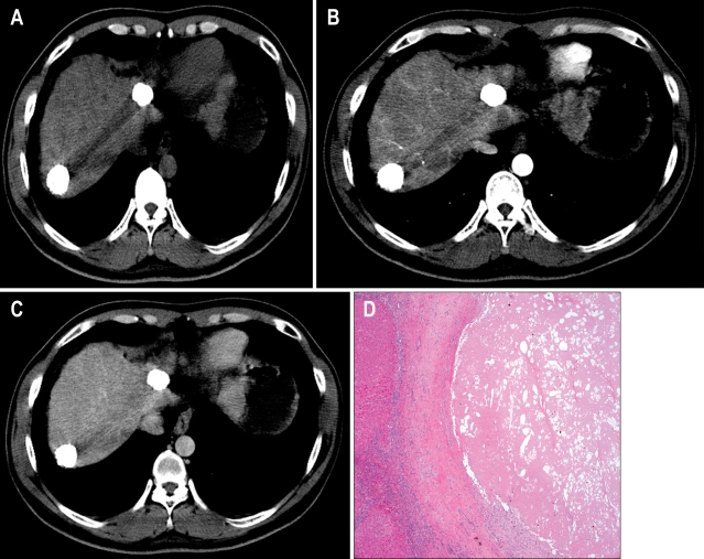
Fig. 1.
Two hepatocellular carcinoma (HCC) nodules with a radiologic response of 39.1 months showing complete necrosis in the explanted liver. (A-C) Computed tomography (CT) showing HCC nodules of 2.5 cm and 2.6 cm with complete lipiodol uptake without enhancement (A, precontrast imaging; B, arterial phase; C, delayed phase). (D) Total tumor necrosis was observed in the explanted liver (H&E stain, ×50).

