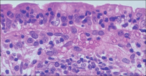
Fig. 2.
High-magnification photograph of the field shown in Fig. 1 showing increased numbers of intraepithelial lymphocytes (Adapted from Freeman HJ. Pearls and pitfalls in the diagnosis of adult celiac disease. Can J Gastroenterol 2008; 22:273-280).

