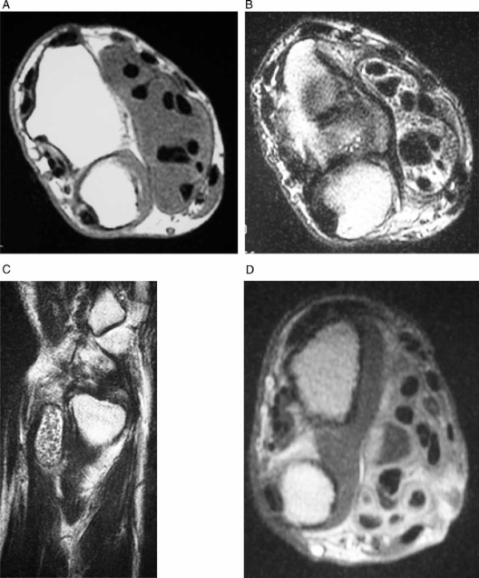
Figure 2.
A: Axial T1-weighted MR image of the right distal forearm demonstrates a mass with low signal intensity surrounding the flexor tendons. B: Axial T2-weighted MR image showed a mass with high signal intensity mass and many rice bodies with low signal intensity. C: Sagittal T2-weighted MR image of the right distal forearm showed enlarged tendon sheaths and many small bodies with low signal intensity. D: Axial T1-weighted MR images of the right distal forearm after the administration of gadolinium. The mass which surrounded the flexor tendons was moderately enhanced.

