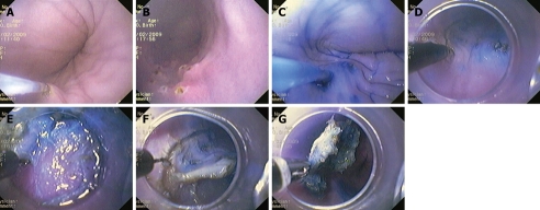
Figure 1.
Procedure. A, B: Artificial lesion marked with coagulation points; C: Injection of normal saline with epinephrine and indigo carmine; D: Knife cutting of a circumference around the lesion; E: Transparent softcap provides lesion counter-traction; F: Soft cap attached to the tip of the endoscope during endoscopic submucosal dissection (ESD); G: Grasping forceps during retrieval of ESD specimens.

