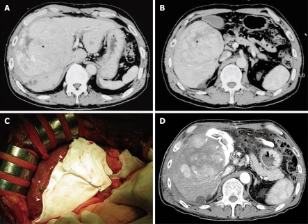
Figure 1.
A huge tumor occupying a wide area of the right lobe. A: Abdominal computed tomography (CT) showing a pre-treatment dominant lesion located widely in the right lobe (asterisk); B: The tumor (asterisk) has extended contact with the gastrointestinal tract; C: Intraoperatively, Gore-Tex sheets (arrowheads) maintained a space between the tumor (asterisk) and the gastrointestinal tract (under the fingertips); D: Post-operative abdominal CT showing the spacer (arrowheads) around the tumor (asterisk); the spacer maintained a sufficient open space between the tumor and the gastrointestinal tract.

