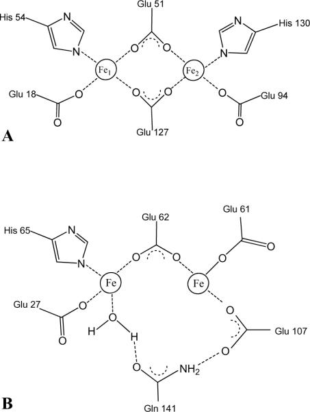
Figure 1.
Schematic representation of: (A) The symmetrical ferroxidase center typical of bacterioferritin. Bridging glutamates are Glu51 and Glu127 and the capping residue pairs are Glu18/His54 and Glu94/His130. (B) The ferroxidase center of human H-chain ferritin adapted from the crystal structure of the Tb3+ derivative (43).

