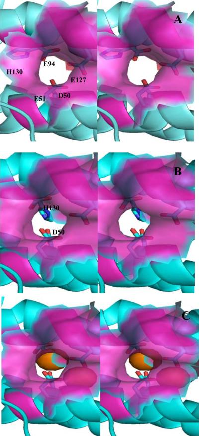
Figure 7.
Stereo view of the ferroxidase pore viewed from the protein exterior. The semi-transparent surface representation (magenta) is constituted by residues lining the pore wall (Asn17, Ile20, Leu93, Lys93 and Ala97). (A) The “gate-open” conformation of the Asp50 and His130 side chains (observed in the as isolated and mineralized Pa BfrB structures) allows a nearly unobstructed view of the interior cavity through the pore. (B) The “gate-closed” conformation of Asp50 and His130 (observed in the Fe-soaked structure) obstruct the bottom of the pore and poise H130 to coordinate Fe2. The ferroxidase iron is not shown for clarity. (C) View identical as in (B) but showing the ferroxidase iron ions as orange spheres. The side chain is His130 is below Fe2, which is located at the bottom of the pore. Fe1 in the interior can be seen through the semi-transparent surface.

