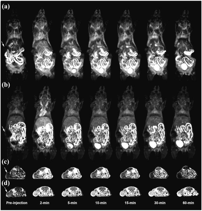
Figure 1.
3D maximum intensity projection images for G2 (a) and CLT1 peptide targeted G2 (b), 2D axial T1-weighted spin-echo images of tumor tissue of the G2 (c) and peptide targeted G2 (d) nanoglobular MRI contrast agents intravenously administered at 0.03 mmol-Gd/kg in nu/nu female nude mice bearing MDA MB-231 tumor xenografts. Arrows indicate tumor.

