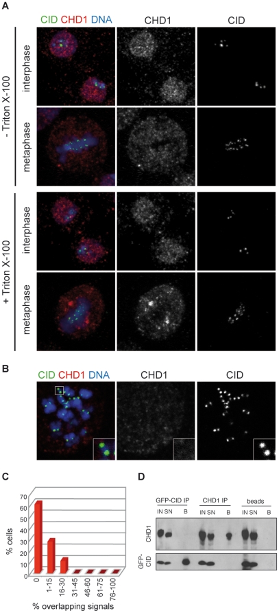
Figure 1. CHD1 is not present at centromeres in Drosophila S2 cells.
A) CHD1 displays nuclear staining during interphase and redistributes to the cytoplasm during mitosis. S2 cells stably expressing EGFP-tagged CenH3CID were treated (bottom) or not (top) with Triton X-100 before fixation to reduce the amount of soluble protein. Cells were stained with anti-CHD1 (red) and anti-GFP (green) antibodies. DNA is shown in blue. B) CHD1 is absent from chromosomes at metaphase. Spreads of metaphase chromosomes from EGFP-CenH3CID-expressing S2 cells were stained with antibodies against CHD1 (red) and GFP (green). C) Quantification of overlapping CHD1 and EGFP-CenH3CID signals. Percentages of overlapping signals per cell were calculated and plotted against the percentage of cells displaying similar ratios of overlap. D) CHD1 and EGFP-CenH3CID do not interact. Co-immunoprecipitations were performed on micrococcal nuclease treated S2 cell extracts with antibodies against GFP, CHD1 or protein A sepharose beads only. Aliquots of the input (IN) fraction, supernatant (SN) and eluted beads (B) were subjected to immunoblotting with anti-CID and anti-CHD1 antibodies.

