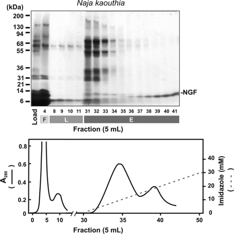
FIGURE 1.
Ni2+-Agarose chromatography of N. kaouthia venom. Ni2+-Agarose chromatography of venom (0.1 mg/10 ml TS buffer) analyzed by SDS 5–20% polyacrylamide gel electrophoresis (reducing conditions) and Coomassie Blue staining is shown, demonstrating load, flow-through (F), lag (L), and eluted (E) fractions. The A280 elution profile (solid line) and the linear 0–30 mm imidazole gradient (dashed line) are shown in the lower panel.

