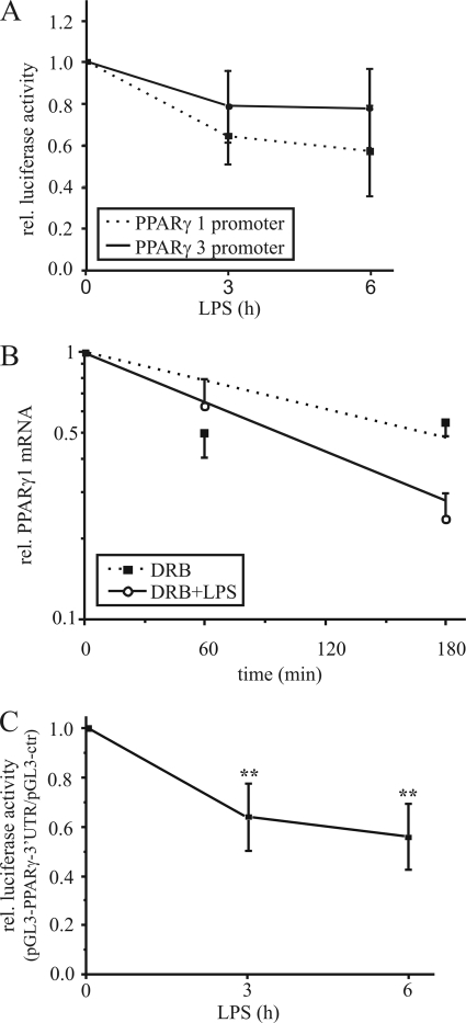
FIGURE 2.
Destabilization of PPARγ1 mRNA. A, PPARγ promoter 1 and 3 activities were determined by reporter assay in THP-1 macrophages, transfected with the promoter constructs and stimulated with 1 μg/ml LPS for 3 and 6 h. B, primary human macrophages were exposed to 100 μm DRB (filled squares) or 1 μg/ml LPS plus DRB (open circles) up to 3 h and total PPARγ1 mRNA (including transcripts 1 and 3) was determined by qPCR. C, differentiated THP-1 cells were transfected with the pGL3-control or pGL3-PPARγ-3′-UTR vector, and reporter activity was analyzed in response to 1 μg/ml LPS. Luciferase activity was normalized to protein and the ratio of pGL3-PPARγ-3′-UTR activity/pGL3-control is displayed. Data present mean values ± S.E., n ≥ 4. Statistics were analyzed with the unpaired Student's t test.*, p < 0.05; **, p < 0.01.

