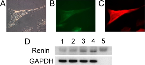
FIGURE 7.
Immunofluorescent staining for α-smooth muscle actin and immunoblotting for renin. A–C, murine MSCs overexpressing GFP-LXRα (mMSC/LXRα/GFP) were treated with cAMP for 5 weeks. A, microscopic findings showed granules in cytoplasm. B, cells induced from mMSC/LXRα/GFP exhibited green fluorescence. C, granulated cells were positive for α-smooth muscle actin. Original magnification, ×400. D, murine MSCs overexpressing GFP alone (mMSC/GFP) and murine MSCs overexpressing GFP-LXRα (mMSC/LXRα/GFP) were treated with or without cAMP daily for 8 weeks. Cell lysates were isolated and subjected to immunoblotting analysis using antibodies for renin and GAPDH. Lane 1, mMSC/GFP without treatment (Tx); lane 2, mMSC/GFP with cAMP Tx; lane 3, mMSC/LXRα/GFP without Tx; lane 4, mMSC/LXRα/GFP with cAMP Tx; lane 5, positive control (human renin recombinant protein).

