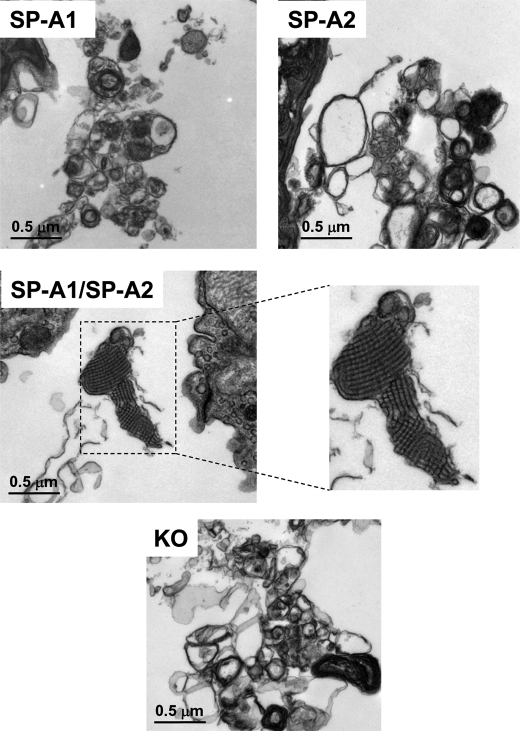
FIGURE 10.
Ultrastructures of the lung tissues from in vivo TM rescue in SP-A KO mice. SP-A KO mice that lack TM structures were treated with exogenous SP-A protein. Fifty μl of saline buffer containing 2.5 or 5 μg of SP-A (one of the three types of SP-A (SP-A1, SP-A2, or SP-A1/SP-A2) described in the legend to Fig. 9) was administered into the lung of SP-KO mouse intratracheally. One negative control used 50 μl of saline without SP-A (see “Materials and Methods”). The mice were killed at 6 and 12 h after treatment, and the lung tissues were fixed immediately with Karnovsky's fixative solution for 24 h. The fixed lung tissues were used for analysis by electron microscopy. Ultrastructures of each sample (5 μg of SP-A and 6 h after treatment) were shown at a magnification of ×30,000. TM structures were observed in the lung tissues from the SP-A KO mice treated with SP-A containing both SP-A1 and SP-A2 gene products but not in those treated with SP-A1 or SP-A2 alone. As expected, no TM structure was found in the negative control.

