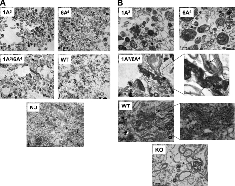
FIGURE 8.
Ultrastructures of surfactant large aggregates from BAL of hTG mice. BAL fluid was obtained from hTG mice with a single gene, either SP-A1(6A4) or SP-A2(1A3), and both genes SP-A1/SP-A2(6A4/1A3) as well as from the same background SP-A KO and WT mice. To generate a sufficient amount of the large aggregate fraction for this assay, BAL fluids from five mice were pooled for each type of samples. Large aggregates were prepared from BAL using sucrose gradient centrifugation. The large aggregate fraction of surfactant was collected from the interface following centrifugation. Pellets of large aggregates were fixed with 2.5% paraformaldehyde and 4% glutaraldehyde. After 24 h, the fixed pellets were used for analysis by electron microscopy. A and B represent ultrastructures of each sample at magnifications of ×10,000 (A) and ×30,000 (B), respectively. In the single gene-containing hTG mice, SP-A1(6A4) and SP-A2(1A3), the lamellar bodies in the large aggregates appear more dense compared with KO mice (A), and tubular myelin figures are absent compared with WT mice (B). hTG mice SP-A1/SP-A2(6A4/1A3) carrying both genes have a similar pattern of ultrastructures of large aggregates as WT mice, including formation of a significant amount of tubular myelin figures marked by arrows (B) in the enlargement of the 1A3/6A4 and WT.

