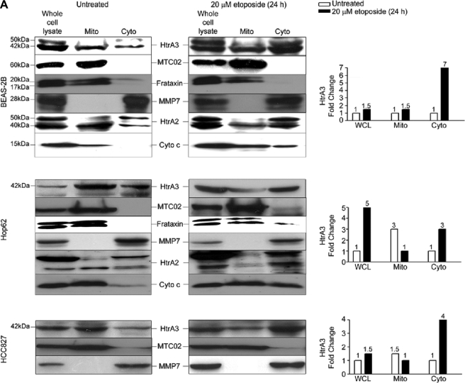
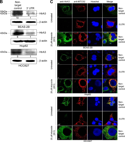
FIGURE 4.
Endogenous HtrA3 is localized to mitochondria and is translocated from mitochondria by etoposide-induced cytotoxic stress. A, immunoblots showing endogenous expression of HtrA3, mitochondrial marker MTC02, human frataxin, MMP7, HtrA2, and cytochrome c in whole cell lysates, mitochondria-enriched fractions (Mito), and postmitochondrial cytoplasmic fractions (Cyto) before and after etoposide treatment in the human bronchial cell line BEAS-2B and in lung cancer cell lines Hop62 and HCC827. Both the mature (40 kDa) and immature (50 kDa) HtrA2 forms are shown. Densitometric analysis shows fold changes in HtrA3 intensity for each cell line. B, immunoblots showing endogenous HtrA3 expression, stable HtrA3 down-regulation by 3′ UTR-targeted shRNA, and β-actin loading controls in pooled stable clonal cell lines. A polyclonal antibody was used for detection of endogenous HtrA3. Densitometric analysis of fold changes in HtrA3 intensity normalized by β-actin loading controls is given for each cell line. C, immunocytochemistry using an HtrA3 polyclonal antibody showing HtrA3 and MTC02 co-localization before and after etoposide-mediated apoptotic stimulation. Hoechst stain binds nuclear material. Original magnification, ×100.


