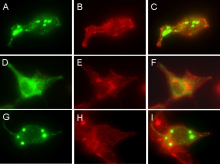
FIGURE 2.
Localizations of full-length, C terminus-deleted, and N terminus-deleted Hv1. HeLa cells grown on glass coverslips were transfected with the expression plasmids, pHv1-EGFP, pΔC-Hv1-EGFP, and pΔN-Hv1-EGFP, and observed under a DM RXA2 microscope. The localizations of full-length (A), C terminus-deleted (D), and N terminus-deleted (G) Hv1 are shown in green. The cytoskeleton is labeled with rhodamine phalloidin (shown in red; B, E, and H). Images merging actin (red) with the full-length Hv1 (C), C terminus-deleted Hv1 (F), and N terminus-deleted Hv1 (I) (green) show that the full-length, C terminus-deleted, and N terminus-deleted Hv1 all localize in intracellular sites, in which the C terminus-deleted Hv1 is present over the entire cell.

