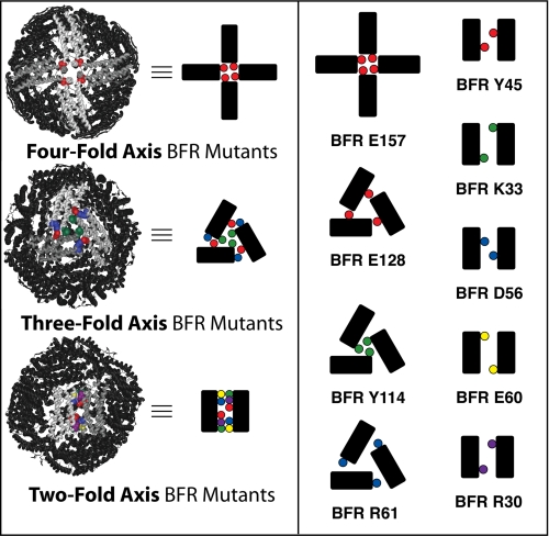
FIGURE 1.
Position of mutated residues with respect to symmetry axes of the nano-cage protein. Left, the BFR crystal structure with mutated residues highlighted with respect to the three axes of symmetry and schematized diagrams representing the protein-protein interactions defining these symmetries. Right, the residues of BFR mutated in this study and their schematized position at the symmetry-related protein-protein interfaces. This schematic convention will be conserved throughout this report.

