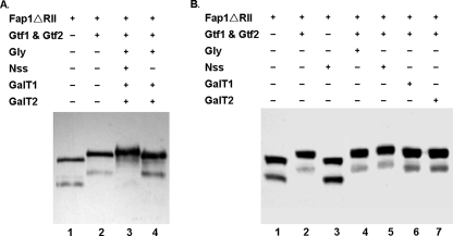
FIGURE 3.
Analysis of Fap1ΔRII expression in a recombinant E. coli glycosylation system. A, an E. coli strain carrying pHSG576/fap1ΔRII was transformed with pGEX-6p-1 (lane 1), pGEX-6p-1/gtf1–2 (lane 2), pGEX-6p-1/gtf1–2/pVPT-gly-nss-galT1-galT2 (lane 3), and pGEX-6p-1/gtf1–2/pVPT-gly-galT1-galT2 (lane 4), respectively, and subjected to Western blotting analysis. B, an E. coli strain carrying pHSG576/fap1ΔRII was transformed with pGEX-6p-1 (lane 1), pGEX-6p-1/gtf1–2 (lane 2), pGEX-6p-1/pVPT-nss (lane 3), pGEX-6p-1/gtf1–2/pVPT-gly (lane 4), pGEX-6p-1/gtf1–2/pVPT-nss (lane 5), pGEX-6p-1/gtf1–2/pVPT-galT1 (lane 6), and pGEX-6p-1/gtf1–2/pVPT-galT2 (lane 7), respectively, and subjected to Western blotting analysis.

