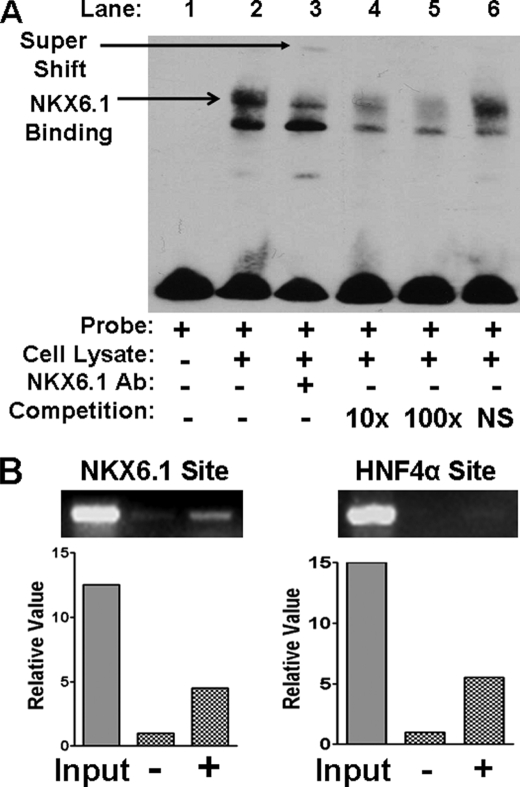
FIGURE 5.
NKX6.1 binding to the HNF1α promoter by EMSA and ChIP. A, EMSA. EMSAs were conducted with INS1 cell lysate and biotin-labeled HNF1α promoter oligonucleotide (Probe) as indicated. Arrows indicate binding and supershift. In competition assays, DNA binding reactions were preincubated with 10-fold (10×) or 100-fold (100×) unlabeled HNF1α oligonucleotide or nonspecific (NS) oligonucleotide as indicated. Ab, antibody. B, ChIP assays. NKX6.1 binding experiments are shown in the left panel with (+) or without (−) NKX6.1 overexpression. HNF4α binding experiments are shown in the right panel, with (+) or without (−) HNF4α overexpression. Representative images are shown following agarose gel electrophoresis of PCR products. Quantification of bands was done with the Microsoft Photoshop quantification tool.

