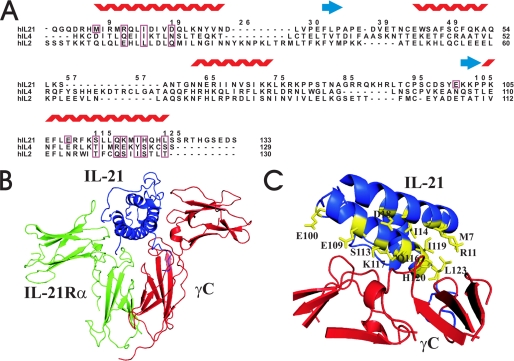
FIGURE 1.
A, sequence alignment of hIL-21, hIL-2, and hIL-4 based on the structural alignment and adjusted by hand. γC binding residues on IL-21 predicted from alignment with IL-2 and IL-4 receptor complexes are boxed in the alignment together with the corresponding residues in IL-2 and IL-4. Three additional γC binding residues identified from the model (M7, E100, E109) are only boxed in the sequence of hIL-21. The numbering follows hIL-21. B, homology model of the IL-21/IL-21Rα/γC complex generated by the Modeler program. The structures of IL-21, IL-21Rα, and γC are depicted as blue, green, and red ribbons, respectively. C, potential γC binding residues of hIL-21 are depicted as yellow sticks.

