FIGURE 2.
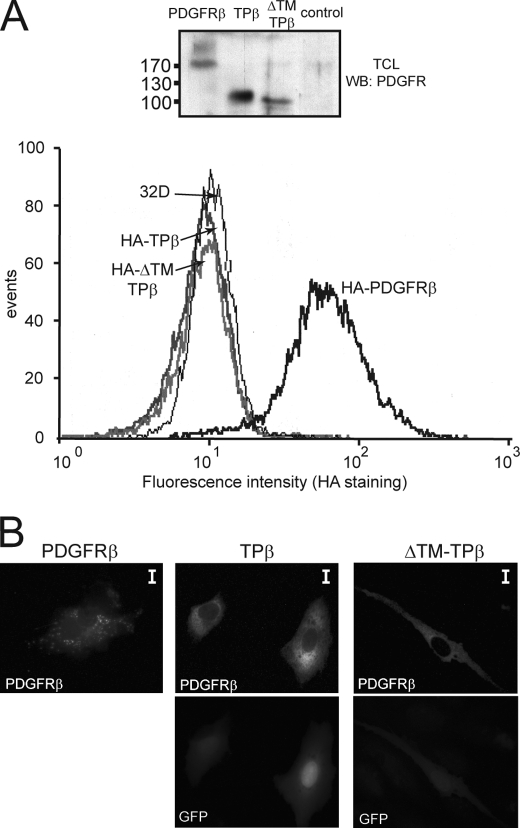
TPβ is not a membrane spanning protein. A, intact 32D cells stably expressing the HA-tagged form of wild-type PDGFRβ, TPβ, or ΔTM-TPβ were stained with anti-HA antibodies and analyzed by flow cytometry. Untransfected 32D cells were used as control. Total cell lysates (TCL) from the same cell lines were analyzed by Western blot (WB) with anti-PDGFRβ antibodies. B, PAE cells were transfected with the indicated receptors and stained with anti-PDGFRβ antibodies and fluorescent secondary antibodies. The cells were analyzed by fluorescent microscopy. GFP is co-expressed with TPβ and ΔTM-TPβ from the bicistronic vector used for transfection. The scale bars correspond to 10 μm.

