FIGURE 2.
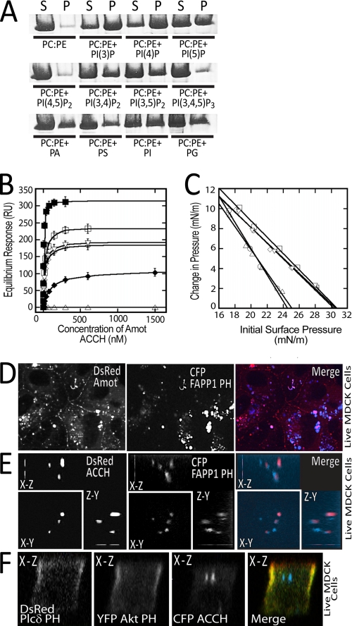
The ACCH domain selectively binds and penetrates membranes containing monophosphorylated phosphatidylinositols. A, the purified human Amot ACCH domain was incubated with liposomes of the indicated compositions (5% PIs or 20% PA, PS, PI, or PG). The supernatant (S) and pellet (P) following sedimentation was resolved by SDS-PAGE and visualized by Coomassie dye. B, the saturation responses (Req) (see supplemental Fig. S2B) at each ACCH domain protein concentration from SPR sensorgrams were plotted versus protein concentration for different liposomes (PM + PI(4)P (■), PM (□), POPC/POPE/PI4P (75:20:5) (▿), POPC/POPE/PI(3)P (75:20:5) (◇), POPC/POPE/PI(5)P (75:20:5) (♦), and POPC/POPE/PI(4,5)P2 (75:20:5) (▵)). The Kd was determined for a minimum of seven different protein concentrations by nonlinear least squares analysis of the binding isotherm using the equation, Req = Rmax/(1 + Kd/C). At least three replicates were done to calculate an S.D. value. C, insertion of the ACCH domain into POPC/POPE (80:20) (○), PM (□), POPC/POPE/PI(4)P (75:20:5) (▿), POPC/POPE/PI(3)P (75:20:5) (◇), and POPC/POPE/PI(4,5)P2 (75:20:5) (▵) monolayers as a function of π. 10 μg of ACCH was injected into the subphase of a monolayer of the indicated initial surface pressure. The Δπ was then monitored for 45 min to construct the curves and determine πc, the x intercept. D, MDCK cells stably expressing DsRed Amot80 and transiently expressing CFP-tagged FAPP1-PH were imaged live. The merge of the Amot (red) and FAPP1-PH (blue) is depicted on right. E, three-dimensional reconstructions of the x-y, x-z, and y-z dimensions of a live MDCK cell grown on a transwell filter expressing Amot ACCH (red) and CFP FAPP1-PH (blue). F, MDCK cells were transfected with vectors that express citrine Akt-PH (green, PIP3 probe), DsRed PLCδ PH (red, PIP2 probe) and cerulean Amot ACCH (blue) (left). Cells were then cultured on a transwell filter for 48 h and then imaged live. The x-z cut section from reconstructed confocal images is shown. mN, millinewtons.

