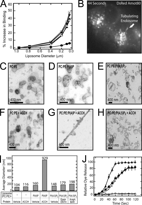
FIGURE 4.
The ACCH domain selectively binds curved membranes and tubulates liposomes containing PI(4)P. A, liposomes with the indicated diameters were analyzed by SPR as described in the legend to Fig. 2 for binding of 100 nm Amot ACCH to POPC/POPE/PI(4)P (75:20:5) (▿), PM (□), POPC/POPE/POPS (60:20:20) (▴), and POPC/POPE/PI (60:20:20) (▾). Also, binding of 100 nm epsin ENTH was monitored to POPC/POPE/PI(4,5)P2 (75:20:5) (●) liposomes of the indicated diameters. B, MDCK cells that stably express DsRed Amot80 were imaged live at 2-s intervals for 80 s. A single frame from this sequence shows Amot marking highly dynamic and often tubulating endosomes (for the complete sequence, see supplemental Movie 2). C–H, liposome samples (1 mg/ml) prepared by extrusion through 100-nm membranes in POPC/POPE (80:20) (C), POPC/POPE/PI(4)P (75:20:5) (D), and POPC/POPE/PI(4,5)P2 (E) were applied to carbon-Formvar-coated copper grids (EMS), and membrane morphologies were examined on an FEI Magellan scanning electron microscope at an electron energy of 15 kV. Representative images were taken with a direct magnification of ×72,000–202,000. The scale bar in each panel is 400 nm. To assess the ability of the ACCH domain to induce lipid curvature changes, liposomes in POPC/POPE (80:20) (F), POPC/POPE/PI(4)P (75:20:5) (G), and POPC/POPE/PI(4,5)P2 (75:20:5) (H) were incubated with 5 μm ACCH domain for 15 min at 25 °C. The sample (8 μl) was then applied to the grids and negatively stained and imaged as described above. I, the size distributions of base POPC/POPE (80:20) liposomes with the indicated lipid ligand (POPC/POPE/PI(4)P (75:20:5) or POPC/POPE/PI(4,5)P2 (75:20:5) were prepared by 100-nm extrusions and assessed by dynamic light scattering analysis. Subsequently, liposomes of the indicated composition were incubated with the respective membrane-tubulating proteins. Reactions were carried to saturation, and then the average diameter of the membranes in each sample was determined by dynamic light scattering analysis. Each bar represents the mean diameter for the respective lipid. J, leakage of 5-carboxyfluorescein dye from liposomes of the indicated compositions by the addition of Amot ACCH protein (POPC/POPE (○), POPC/POPE/PI(4)P (▿), and POPC/POPE/PI(4,5)P2 (▵)) or ENTH domain (POPC/POPE/POPI(4,5)P2 (●)). Triton X-100 disruption of vesicles and subsequent leakage was used as a positive control (▴). At least three replicates were done to calculate an S.D. value for each time point.

