FIGURE 5.
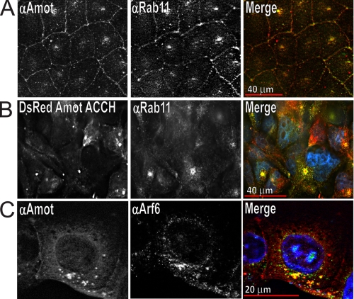
Amot and Amot ACCH similarly distribute in MDCK cells at perinuclear endosomes with Rab11 and Arf6. A, MDCK cells were plated for 18 h, fixed with paraformaldehyde, and immunostained with a rabbit polyclonal antibody directed against Amot (left) and mouse monoclonal antibody directed against Rab-11 (middle) and DAPI stain to visualize nuclei. The merger of Amot (red), Rab11 (green), and DAPI stain (blue) is shown on the right. B, MDCK cells stably expressing DsRed Amot ACCH were plated for 24 h before being fixed with paraformaldehyde and immunostained for Rab11 and stained with DAPI. Left, DsRed Amot ACCH; middle, Rab11; right, merge of Amot (red), Rab11 (green), and DAPI (blue). C, MDCK cells were plated for 18 h and then immunostained for endogenous Amot (left) and Arf-6 (middle). The merger of Amot (red), Arf6 (green), and DAPI (blue) is shown on the right.

