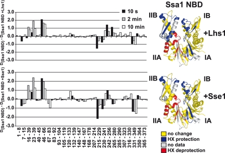
FIGURE 6.
Lhs1 and Sse1 use the same NEF mechanism. Difference in deuteron incorporation between monomeric Ssa1 NBD and NBD in complex with Lhs1 (upper left panel) or Sse1 (lower left panel). The data were resolved to individual non-redundant peptic peptides as indicated by the start and end residue numbers of the corresponding segments. The data presented are from data set two (see supplemental Fig. S2, C and D). Structural representations of the Ssa1 NBD are colored to display the Lhs1 (upper right panel) and Sse1 (lower right panel) induced changes as in Fig. 5B.

