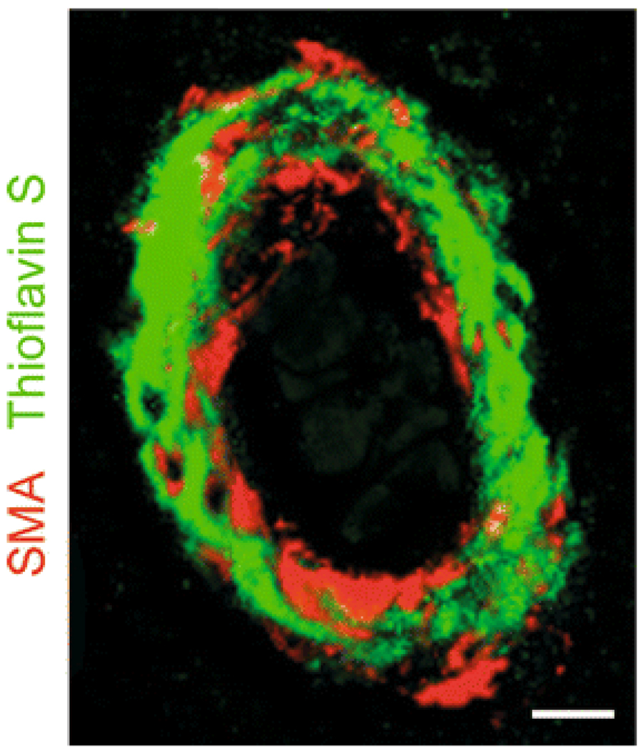
Fig. 1.
Cerebral amyloid angiopathy in AD. Immunofluorescent staining of smooth muscle α actin (SMA; red) and amyloid staining (thioflavin S, green) in an AD cerebral vessel from Brodmann area 9. Staining shows significant amyloid accumulation in the vascular smooth muscle cell (VSMC) layer of this blood vessel. Amyloid accumulation may result from decreased Aβ clearance along the perivascular spaces caused by a decreased low-density lipoprotein receptor related protein-1 (LRP)-mediated Aβ clearance by VSMC, faulty Aβ clearance by perivascular macrophages and/or reduced passive Aβ drainage due to reductions in the arterial pulsatile blood flow. CAA can lead to spontaneous hemorrhage and rupture of the vessel wall due to a loss of the VSMC layer, enzymatically-induced breakdown of the vessel wall, oxidant stress and cytokine-mediated vascular injury. Scale bar 25 µm

