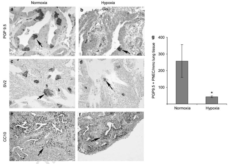
Figure 5.

Immunohistochemical labeling of PNEC/NEB cells (a–d) and Clara cells (e, f) in E12 lung organ cultures maintained in normoxia or hypoxia for 6 days. In normoxia cultures (a, c), the size and number of PNEC/NEB cells (arrow) immunostained for PGP9.5 and neuroendocrine marker SV2 were markedly increased compared with that of cultures maintained in hypoxia (b, d) (original magnification × 400). Expression of Clara cell marker CC10 in airway epithelial cells (arrow) was comparable between cultures exposed to normoxia (e) or hypoxia (f) (original magnification × 200). (g) Quantification of immunoreactive PNEC/NEB cells per mm2 of lung tissue (n = 5) confirmed significant reduction (*P < 0.05) in these cells in lung explants maintained under hypoxia compared with those under normoxia.
