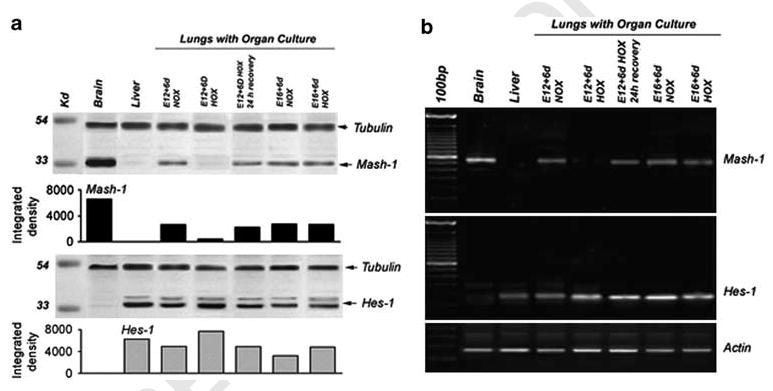
Figure 8.

Analysis for expression of Mash-1 and Hes-1 in lung organ cultures. Western blot (a) and RT-PCR (b) were performed on E12 and E16 lungs in organ culture under normoxia and hypoxia. E12 lungs cultured under normoxia express Mash-1, whereas E12 lungs cultured under hypoxia show almost complete loss of Mash-1 expression. However, when these hypoxia-cultured lung explants were returned to normoxia for 24 h, there was a significant upregulation in Mash-1 expression (recovery). In contrast, E16 lungs did not exhibit any loss of Mash-1 expression under hypoxia. In comparison, Hes-1 expression was maintained under all conditions and gestational ages. Densitometric analysis of western blots shown indicates large differences for Mash-1 and some variance for Hes-1, all relative to positive controls for Mash-1 (brain) and Hes-1 (liver). Loading controls are tubulin for western blots and actin for RT-PCR.
