Fig 2.

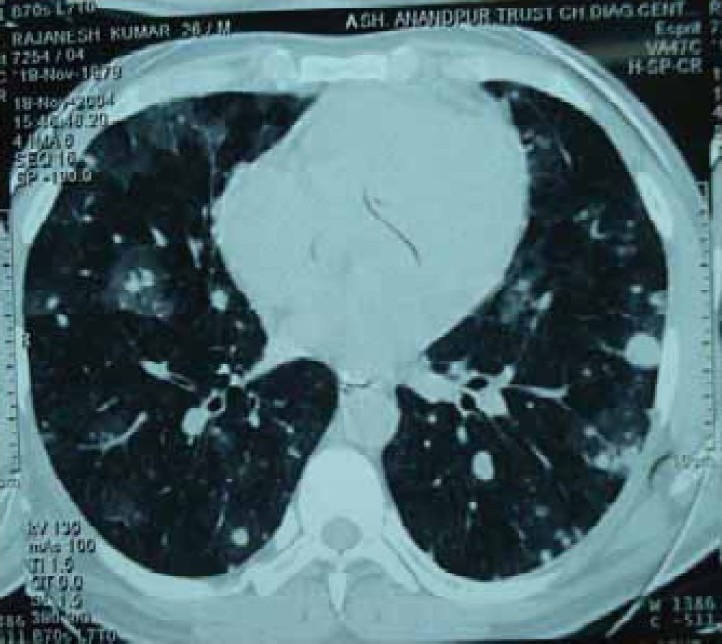
Computed tomographic scan of chest showing multiple patchy ground glass opacities bilaterally with normal intervening lung parenchyma and small nodular opacities scattered in subpleural area

Computed tomographic scan of chest showing multiple patchy ground glass opacities bilaterally with normal intervening lung parenchyma and small nodular opacities scattered in subpleural area