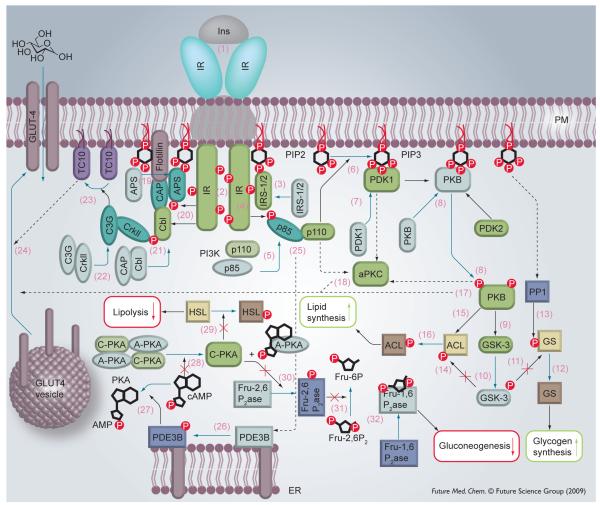
Figure 1. Insulin signal transduction.
Inactive proteins are indicated by a paler color. Protein shape and color depend on the protein function: kinases (and insulin receptor substrates) are green and rectangular with rounded edges. Regulatory domains (e.g., the p85 domain of PI3 kinase) and adaptor proteins are light blue and oval. Phosphatases are dark blue and rectangular. Lyases and synthases are brown. Blue arrows denote a chemical transformation (e.g., PIP2→PIP3) as well as movement of proteins. Black arrows point to the protein target. A solid arrow indicates that the reaction has been established. Dashed arrows refer to pathways that are not yet well established.

