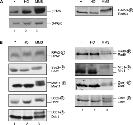
Figure 4.
Histone H2A and RPA2 are phosphorylated in G1-arrested cells after induction of a single DSB. (A) Cultures of wild-type cells (WDHY2172) were arrested in G1 and left untreated (lanes 1), HO-endonuclease was induced (lanes 2) or 0.1% MMS (lanes 3) was added for 2 h. Levels of phosphorylated histone H2A (γ-H2AX) were determined using a modification-specific antibody and quantified against the 3-PGK loading control by densitometry. One example of the anti-Rad53 immunoblots of the corresponding samples in A and B is shown, and in all cases tested Rad53 was induced by MMS and not induced by expression of HO. (B) Cultures of wild-type cells (WDHY2172) or cells with HA-tagged versions of Ddc1 (WDHY2460), Sae2 (WDHY2462), Mre11 (WDHY2502), Ddc2 (WDHY2517), or Chk1 (YLL839) or a Myc-tagged version of Mrc1 (WDHY2180) were treated as in (A). The indicated proteins were analyzed by immunoblotting in total cell extracts using suitable antibodies (see ‘Materials and Methods’ section). Electrophoretic mobility retardation indicates phosphorylation of the corresponding protein.

