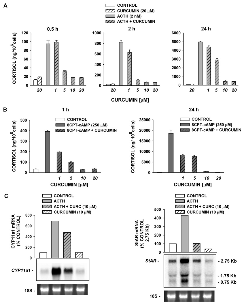
Figure 2.
Curcumin inhibits ACTH- and 8CPT-cAMP-mediated effects on AZF cells. (A) Bovine AZF cells were treated either without (control, white bar) or with curcumin (20 µM, striped bar), ACTH (gray bar), or ACTH plus curcumin (1–20 µM, gray striped bars) for 0.5, 2, and 24 h, as indicated. (B) Cells were treated either without (control, white bar) or with 8CPT-cAMP alone (250 µM, dark gray bar) or 8CPT-cAMP plus curcumin (1–20 µM, dark gray striped bars). Media was collected after 2 h (left panel) or 24 h (right panel) and cortisol concentration determined by EIA as described in the Experimental Section. Cortisol values expressed are the mean ± SEM of duplicates from duplicate samples. (C) Total AZF cell RNA was isolated after 24 h, electrophoresed, blotted, and probed as described in the Experimental Section. Each lane contained 10 µg of total RNA. Membranes were probed with specific probe for bovine CYP11a1 (left) or bovine StAR (right). Cells were incubated either without (control, white bar), with ACTH alone (2 nM, gray bar), or ACTH (2 nM) plus curcumin (10 µM, gray striped bar), or curcumin alone (white striped bar), as indicated. mRNA is expressed as % control value determined using Kodak 1D 3.6 Image Analysis software. Representative blots from ≥2 different experiments are shown. 18S rRNA bands from gel is shown as evidence of even loading.

