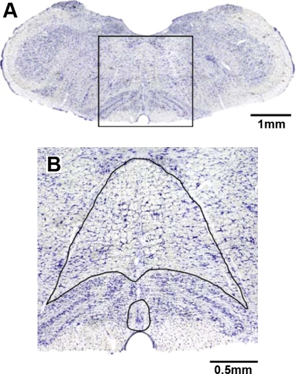
Fig. 1.
Area of the medulla analyzed for total neurons and K+ channel immunoreactive neurons. A: transverse section (1× magnification) at obex from a Dahl salt-sensitive (SS) rat that was stained for Nissl substance. B: the box in A at 10× magnification, wherein the neuron count area is outlined. The small area is raphé pallidus ventral to the olivary nuclei and at the midline between the olivary nuclei. The larger enclosed area is raphé obscurus dorsal to the olivary nucleus and medial and ventral to the hypoglossal nerves.

