Fig. 10.
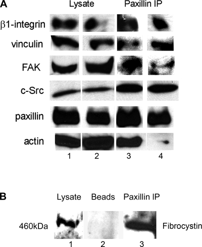
Composition of paxillin-containing focal adhesion complex in ARPKD cells compared with normal HFCT cells. A: Western immunoblot of HFCT (lane 1) and ARPKD (lane 2) cells at steady state. Cell lysate, 30 μg per lane. Anti β1-integrin, 1:500; anti-vinculin, 1:1,000; anti-FAK, 1:500; anti-c-Src 1:1,000; anti-paxillin, 1:2,000; anti-actin, 1:10,000. Identical cells: HFCT (lane 3) and ARPKD (lane 4) cells were immunoprecipitated with anti-paxillin 1 μg/1 mg of lysate and immunoprecipitates (IP) were subjected to immunoblot analysis. Note that very little actin was detected in the paxillin immunoprecipitates from ARPKD cells. B: association between paxillin and fibrocystin-1. Western immunoblot analysis of fibrocystin-1 in 30 μg mouse inner medullary collecting duct (mIMCD) cell lysate (lane 1) and paxillin immunoprecipitate from 1 mg of lysate (lane 3). Lane 2 is bead-only negative control. Anti-paxillin 1:2,000; anti-fibrocystin-1, 1:500.

