Fig. 11.
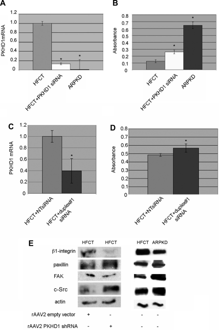
Effects of siRNA or short hairpin RNA (shRNA) knockdown of PKHD1 in normal HFCT cells. A: real-time quantitative PCR (qPCR) of PKHD1 mRNA content of HFCT cells transfected with PKHD1 siRNA, normalized to levels of actin mRNA. Note that PKHD1 siRNA reduced PKHD1 mRNA by 85% (white vs. gray bar). Negligible levels of PKHD1 mRNA were seen in ARPKD cells (black bar); n = 3; Student's t-test, *P < 0.05, compared with HFCT. B: MTS adhesion assay 4 h after plating 2,000 cells on collagen I. HFCT cells transfected with PKHDI siRNA (white bar) showed increased levels of adhesion compared with untreated HFCT (gray bar). Student's t-test, *P < 0.0001, n = 10, compared with HFCT. C: real-time qPCR of PKHD1 mRNA content of HFCT cells transfected with PKHD1 siRNA, duplex#1 compared with nontargeting (NT) sequence. Levels normalized to actin mRNA (n = 3). Student's t-test, *P < 0.05, compared with HFCT + NT. Note that PKHD1 mRNA was reduced 60%. D: MTS adhesion assay 4 h after plating 2,000 cells on collagen I. HFCT cells transfected with PKHDI siRNA duplex#1 showed increased levels of adhesion compared with HFCT cells transfected with NT sequence. Student's t-test, *P < 0.001, n = 10, compared with HFCT +NT. E: Western immunoblot analysis of attachment-dependent expression of focal adhesion complex proteins after rAAV2GFP-PKHD1 shRNA knockdown (>90% reduction PKHD1 mRNA and protein) in HFCT cells. After 24 h transduction with rAAV2GFP-PKHD1shRNA or empty vector (rAAV2-GFP), 5 × 10⁶ cells were plated for 4-h on type I collagen and attached cells collected. Lysate, 15 μg per lane. Anti-β1-integrin, 1:500; anti-paxillin, 1:2,000; anti-FAK, 1:500; anti-c-Src, 1:1,000; anti-actin, 1:10,000.

