Fig. 2.
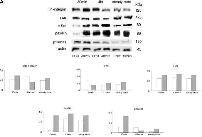
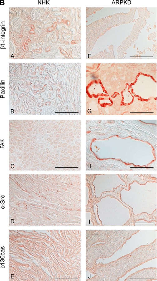
Time- and attachment-dependent expression of total levels of focal adhesion complex proteins in ARPKD and HFCT cells. A: Western immunoblot analysis of focal adhesion proteins in age-matched normal HFCT and ARPKD epithelial cell lines. Equal numbers of cells in suspension were plated on collagen I for 30 min, 4 h, or 7–11 days (confluent steady state); attached cells were analyzed for content of total β1-integrin, focal adhesion kinase (FAK), c-Src, paxillin, and p130cas; and levels were normalized to actin content. Cell lysate, 30 μg per lane. Anti-β1-integrin, 1:500; anti-FAK, 1:500; anti-c-Src, 1:150; anti-paxillin, 1:2,000; anti-p130cas, 1:500; anti-actin, 1:10,000. Densitometry analysis was carried out for focal adhesion proteins at each time point. B: immunohistochemistry of focal adhesion complex proteins in age-matched pediatric (4–5 yr) normal human kidney (NHK; A–E) and end-stage ARPKD (F–J). Anti-β1-integrin, 1:50; anti-paxillin, 1:200; anti-FAK, 1:50; anti-c-Src, 1:150; anti-p130cas, 1:50. Aminoethylcarbazole (red) reaction product. Bright-field and Nomarski optics. Scale bar represents 100 μm.


