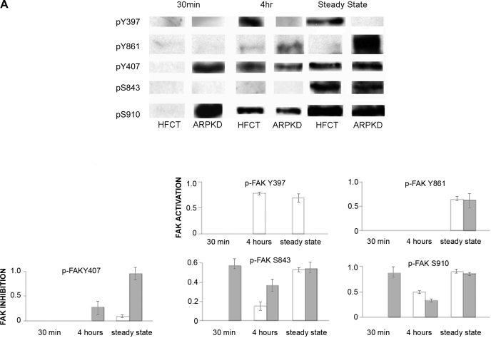
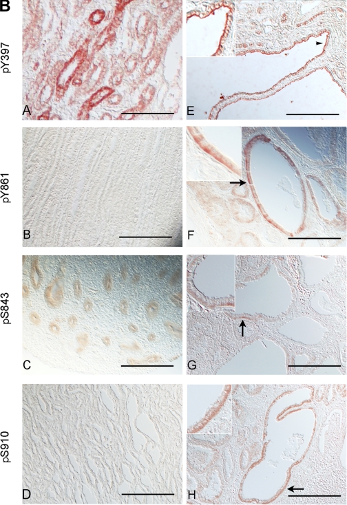
Fig. 3.
Time- and attachment-dependent site-specific phosphorylation of FAK in ARPKD cells by comparison to normal HFCT epithelia. A: Western immunoblot analysis of site-specific phosphorylation of FAK. Equal numbers of cells were plated on collagen I for 30 min, 4 h, or 7–11 days (confluent steady state) and attached cells were analyzed for pY397-FAK, pY407-FAK, pY861-FAK, pS843-FAK, and pS910-FAK. Cell lysate, 45 μg per lane. Anti-pY397-FAK, 1:1,000; anti-PY407-FAK, 1:1,000; anti-pY861-FAK, 1:1,000; anti-pS843-FAK, 1:2,000; anti-pS910-FAK, 1:2,000. After initial immunoblotting, membranes were stripped and reimmunoblotted for total FAK. Densitometry analysis was carried out and the fraction of phosphorylated FAK to total FAK levels were calculated at each time point. B: immunohistochemistry of phospho-FAK in age-matched pediatric (4–5 yr) normal human kidneys (A–D) and end-stage ARPKD (E–H) tissue sections. Anti-pY397-FAK, 1:100; anti-pY861-FAK, 1:100; anti-pS843-FAK, 1:200; anti-pS910-FAK 1:200. Aminoethylcarbazole (red) reaction product. Most pY861-, pS843- and pS910-FAK staining was seen in basal cell membranes in ARPKD (F–H, arrows and insets). By contrast, pY397-FAK was localized to apical membranes of ARPKD cyst epithelia (E, arrowhead and inset). Bright-field and Nomarski optics. Scale bar represents 100 μm.


