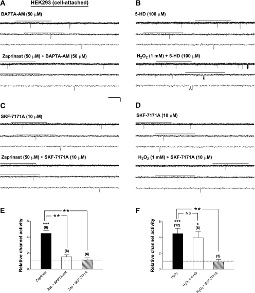
Fig. 6.
Stimulation of Kir6.2/SUR1 channels by PKG activators and exogenous H2O2 in intact cells is sensitive to suppression of intracellular Ca2+/calmodulin activities, whereas the stimulatory effect of exogenous H2O2 on the channel is not affected by the mitoKATP channel inhibitor 5-HD. Currents were obtained in the cell-attached patch configuration from transiently transfected HEK293 cells expressing Kir6.2/SUR1 channels. A–D: single-channel current traces of Kir6.2/SUR1 channels in a cell-attached patch before (top traces) and during (bottom traces) bath perfusion of zaprinast (50 μM; A and C) or H2O2 (1 mM; B and D) in the presence of the membrane-permeable Ca2+ chelator BAPTA-AM (50 μM; A), the mitoKATP channel inhibitor 5-HD (100 μM; B), or the membrane-permeable calmodulin antagonist SKF-7171A (10 μM; C and D). Scale bars are the same as in Fig. 1. E and F: averaged relative single-channel activity (i.e., normalized NPo) of Kir6.2/SUR1 channels obtained during drug application in individual groups. Drug effect on the relative channel activity was compared with the corresponding control obtained from the same patch and normalized as described in Fig. 1F (control as 1, dashed line). Data are means ± SE of 6–12 patches. *P < 0.05; **P < 0.01; ***P < 0.001 (two-tailed, one-sample t-tests within individual groups or unpaired t-tests between groups).

