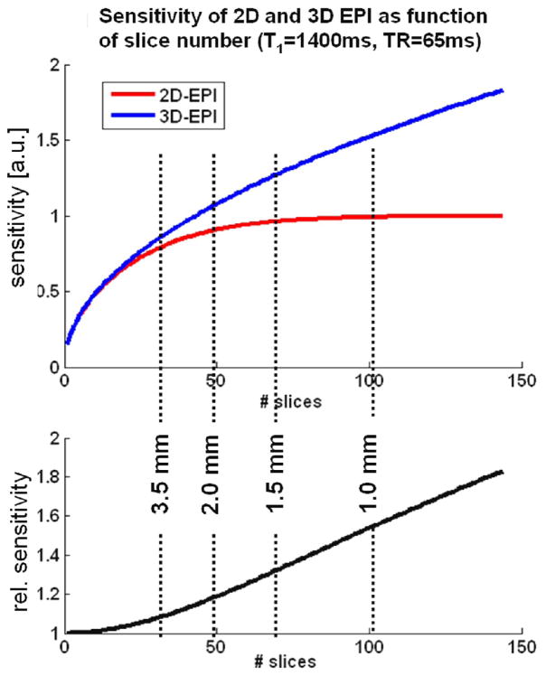
Fig 1.
Theoretical sensitivity of 2D and 3D EPI as a function of the number of acquired slices (top) the relative performance of 3D vs. 2D EPI (bottom) in the absence of physiological noise as derived from the signal equations (Eqs1 and 2) for the case without parallel undersampling. Since the resulting volume TR are identical for both methods, their relative sensitivities scale with the MR signal. The dashed lines indicate the approximate number of slices needed for whole-brain coverage (≥ 100mm axial slab) at the different spatial resolutions.

