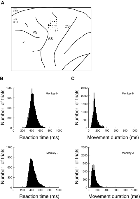
Fig. 1.
A: location of electrode penetrations in dorsal premotor area (PMd) of monkey H. The size of the dots represents the number of neurons recorded. CS, central sulcus; AS, arcuate sulcus; PS, principal sulcus. B: histogram of reach reaction times in the search task. C: histogram of reach durations in the search task.

