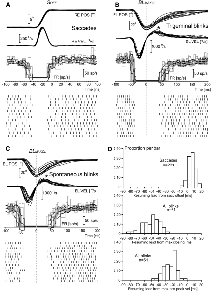
Fig. 4.
OPN resumption leads: single trials. The first 3 panels illustrate movement position and velocity traces, OPN firing frequency, and OPN spike events as a function of time for subsets of single trials from cell c51026.A1, the same cell of Fig. 2. The layout of these 3 panels is identical to that of Fig. 2, with the following difference: the saccadic traces (A) are now synchronized with the offset of the saccade, identified with the vertical dotted line labeled SOFF, and the blink traces are synchronized with respect to the time of maximum downward closing of the eyelid, identified with the vertical dotted line labeled BLMAXCL. The cell resumed around the end of the saccade (A), which is also near when the saccadic short-lead burst neurons end their activity. For both trigeminal (B) and spontaneous (C) blinks, the cell resumed several 10 s of milliseconds after the maximum closing of the eyelid, which human electromyographic (EMG) data indicate it occurs near the end of the activity in the orbicularis oculi motoneurons (OOMNs). This striking difference is also evident in the histograms in D. Positive values mean that the cell resumed before the specific movement event. For the blink histograms, trigeminal and spontaneous data are pooled together. The third histogram illustrates that the OPN resumption occurred even after the time of the maximum positive velocity peak of the reopening phase, indicated by the asterisks in B and C. The human EMG data indicate that this event is reached upto 50 ms after the end of the activity in the orbicularis oculi muscle, as the result of passive elastic forces and the resumption of activity in the levator palpabrae superioris muscle.

